Kenosha Reuse Discussion Board > GURNEE, IL
> Commercial
> Mechanical
> Used
> Zeiss ff 450PLUS ir fundus camera visupac FF450 plus ir
Zeiss ff 450PLUS ir fundus camera visupac FF450 plus ir
(1) PRE-OWNED AND IN EXCELLENT CONDITION
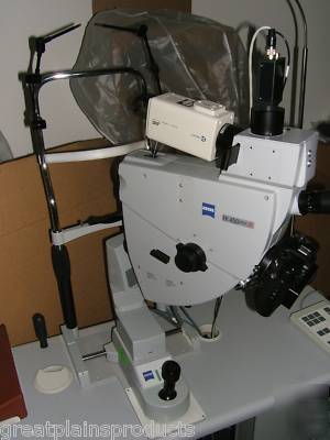
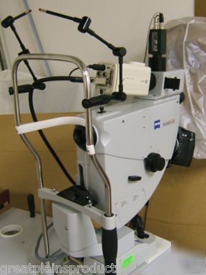
ZEISS VISUPAC And FF450 PLUS IR FUNDUS CAMERA
*The Fundus Camera And VISUPAC Have Been Inspected, Tested AND Passed By ZEISS.
This Equipment Is Ready To Go To Work In Your Clinic Or Practice Today!*
* (1) ZEISS FF450PLUS IR FUNDUS CAMERA
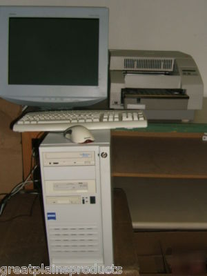
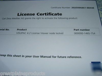
* (1) VISUPAC SYSTEM 4.2.2 (Includes Zeiss License Certificate)
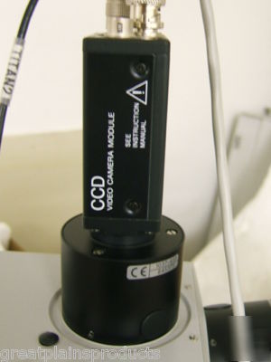
* (1) SONY CCD VIDEO CAMERA MODULE: MODEL# XC-75CE (DC 10.5 - 15V)
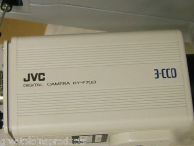
* (1) JVC DIGITAL CAMERA (Model KY-F70BU)
* (1) ZEISS POWER TABLE / POWER STAND
* (1) OPERATOR'S MANUAL (HARD COPY) FOR THE FF450PLUS IR FUNDUS CAMERA
* (1) VISUPAC OPERATOR'S MANUAL (Inside The Software)
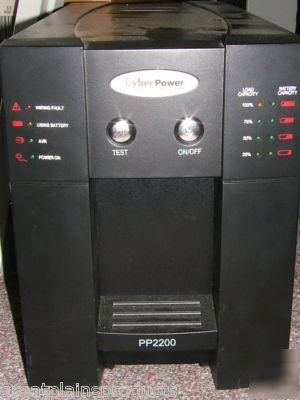
* (1) CYBER POWER PP2200 POWER SUPPLY (Please Note That The Plug-In Does Not Insert Into A Typical Outlet, This Is A 30 Amp Or 50 Amp Plug-In)
* (1) SONY TRINITRON MULTISCAN G520 COLOR MONITOR
* (1) KODAK PROFESSIONAL 8670 PS THERMAL PRINTER (Please Note That There Is Not A Warranty On This Printer)
VISUPAC And FF 450plus Fundus Camera
Working Together For Enhanced Patient Care
* The best diagnosis starts with the best images.
Advanced digital sensor technology provides the sharpest fundus detail available
* Exclusive ZEISS telecentric optics. Proprietary high-transmission, high-resolution optical features
* Integrates seamlessly with the FF 450plus Fundus Camera for superb image quality, diagnostic versatility and upgradeability to future technologies
* Powerful computer/software is easily accessed with a simple, straightforward user interface
* Incorporates the highest-resolution optics and the most advanced digital sensor technology to provide the sharpest fundus detail available
VISUPAC and FF 450plus IR Fundus Camera
The VISUPAC system and the FF 450plus Fundus Camera are reciprocally calibrated, consistently providing a unique level of accuracy and security that can't be approximated with a third-party or aftermarket configuration. Without this level of integrated synergy, calibration and measurement accuracy cannot be assured.
Matching the VISUPAC system pixel for pixel, the FF 450plus Fundus Camera sets the industry standard for image quality, offering outstanding, highly detailed image capture that optimizes the capabilities of the VISUPAC system.
The FF 450plus provides other ideal synergies to the VISUPAC system.
The FF 450plus ensures fast, easy operation with motorized filters and professional exposure adjustment capabilities with comprehensive default settings for every capture mode. It also provides a choice of sensor resolution for the broad range of clinical needs.
Exclusive ZEISS Telecentric Optics
With the VISUPAC system and FF 450plus Fundus Camera, proprietary high-transmission, high-resolution ZEISS telecentric optics neutralize the effect of the patient's emmetropia, eliminating the potential for image distortion and delivering repeatable and precise measurements of retinal physiology. The system also offers excellent light yield with minimal exposure to the patient.
Sophisticated Tools For Learning
plus Fundus Camera include a unique retina atlas module for new photographers. This sophisticated teaching tool provides a wide array of image samples for a variety of disease states, with descriptions of how the images were obtained. As a result, it offers invaluable guidance on detecting and effectively documenting retinal pathology.
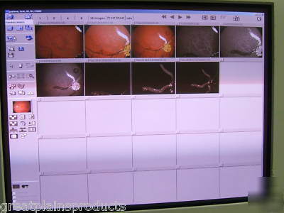
VISUPAC Manages Data So You Can Manage Disease
The digital documentation and image management capabilities of the VISUPAC system were designed for the way ophthalmologists like to work: efficiently, productively and dependably. The VISUPAC system offers an intuitive graphic interface and immediate access to all the information necessary to allow you to confidently diagnose and treat retinal disease. Plus, VISUPAC requires minimal operator training, so it can rapidly take its place in your practice without a significant impact on office workflow.
The VISUPAC system s advanced database includes:
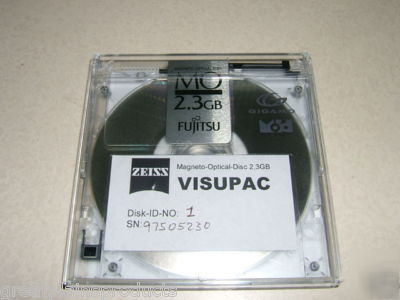
One-Touch Smart Archiving With the touch of a button, all exam information is automatically archived to magneto optical disks (MODs) or your networked server for maximum security and immediate retrieval.
User-Management Engine for HIPAA Compliance:
To meet HIPAA requirements, all patient data is secured to the SQL database structure in individual user accounts, providing safety, privacy and secure access in even the largest LAN/WAN network environments.
Get the complete picture with a quick and comprehensive on-screen review of all previous patient visits.
With the DICOM Imaging Standard, you are able to efficiently retrieve and review patient images and data from virtually any networked workstation, either from a remote location or within your own office; to meet your needs, the VISUPAC system goes beyond simple DICOM compliance to enable the highest standards of workflow, data archiving and network integration for optimal processing and data throughput.
Sophisticated VISUPAC Measurement Modules Include:
Seamless integration with the Stratus OCT system dramatically expands the structural information available to you in support of a thorough and accurate diagnosis.
PDT Calculation (GLD, LSS, BSA)
VISUPAC supports precise PDT preparation by easily enabling determination of greatest linear dimension and laser spot size; the system also automatically calculates body surface area based on patient height and weight and provides a reliable basis for choosing the correct infusion dosage.
Certified Measurement Accuracy:
Retinal measurements within VISUPAC are based on the Gullstrand eye, which can be manually modified to account for corneal radius and refractive error.
VISUPAC provides an intuitive digital framework for documentation of findings for clinical trials per accepted reporting protocols. VISUPAC is accepted by the University of Wisconsin Fundus Photograph Reading Center (UW-FPRC), the Digital Angiography Reading Center (DARC) and the Wilmer Reading Center (WRC).
Cup-to-Disc Ratio, Disc Diameter and Pre-defined Area Measurements:
The VISUPAC system precisely and efficiently calculates all three of these crucial parameters, supporting a more informed and confident diagnosis.
The VISUPAC System Offers A Variety Of Graphic-Based Image Enhancement Features:
Automatic Mapping for Montage (Zeiss Automap)
VISUPAC supports automatic composition of peripheral field montages, or panoramas, by precisely aligning multiple single-field images and matching brightness to provide a complete, wide-angle, highly detailed retinal view.
With this function, clinicians can overlay one image on another to optimize visualization of retinal detail. Image transparency can be varied from zero to 100 percent to better compare pre- and post-visits.
Rapidly view images sequentially and display multiple captured images overlayed to better visualize pathology and make precise diagnoses.
Shapes and text can be drawn on images to more easily locate and identify pathology; modifications can be made at any time, and you are always assured that the original data is never permanently altered once captured.
Additional capabilities include importation of movie, text and PDF files, web browser interface and generation of physician letter templates for detailed and comprehensive patient communication and documentation.
VISUPAC Digital Imaging Archive Management And FF 450
Are an integral - and fully integrated - approach to optimal diagnostic insight and disease monitoring, providing clinical images of unsurpassed quality and an advanced digital imaging and achieve management system that enables unmatched patient care.
made for each other and recipocally calibrated set the industry standard for image quality
deliver repeatable and precise measurements with the exclusive Zeiss telecentic optics
designed for the way ophthalmologists like to work efficiently
easily networked within your existing office
include sophisticated tools for learning and a unique retina atlas module and a wide array of image samples
Integrated System....Optimal Choice
The VISUPAC system incorporates the highest-resolution optics and the most advanced digital sensor technology to provide the sharpest fundus detail available. Plus, with its leading-edge database structure, image manipulation, storage capabilities and data security functions, the VISUPAC system helps ensure optimal diagnostic insight and long-term management of retinal disease.
System Integration For Improved Patient Care
plus Fundus Camera are easily networked within your existing office infrastructure for enhanced efficiency and improved patient care. For instance, both systems can be integrated with the Stratus OCT Optical Coherence Tomographer, allowing you to effortlessly import Stratus OCT scans and data for side-by-side review with VISUPAC images.
Observation Magnification: 11x 19x 29x
Image Scale On 35mm Film: 1,9 2,9 4,3
Frame Format On 35mm Film: 26mm Diameter, Height Limited To 24mm
Visual Observation: Special 10x Eyepiece With Reticle, Monocular
Front Lens To Patient s Eye: 42 mm
Observer s Eye To Patient s Eye: 470 mm
Compensation For Ametropia: +/- 30 D, Continuous
Flash Rate: 1x Per Second (Irrespective Of Flash Level)
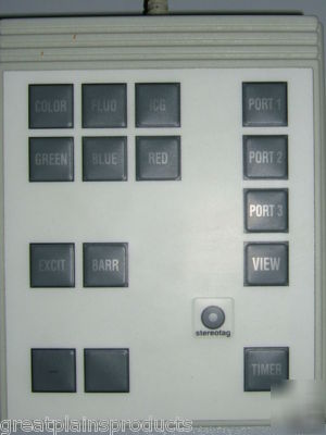
Filters (Motorized Control): Green (RF), Blue, Red Filters, FLUO Filter, ICG Filter (Only Usable In The IR Version), Optional Accessory: Fundus Autofluorescence Filter Set
Special Features: Telecentic Optics, Electronic Interface For VISUPAC Digital Image Archiving Systems, Transmission Of Field angle, OD/OS, Flash Levels
Panning And Tilting Ranges: +/-45 horizontally, +15 /-10 Vertically Using A Hand Wheel
Instrument Table: Asymmetric, Motorized, Suitable For Patients In Wheelchairs, Separate Cable Routing Available For Electronic Sensors
plus Basic System 16.9 x 12.2 x 31.5; 47.4 lb
IT3F Instrument Table 34.7 x 26.2 x max.40.2; 72.8 lb
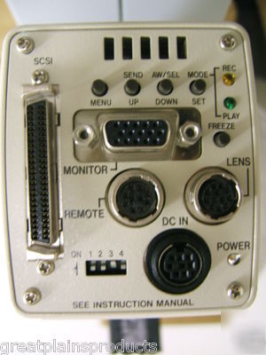
Camera Control Panel 8.9 x 5.9 x 1.8; 2.6 lb
Capture Modes: Color Photography, Red-Free Photography, Red And Blue Pictures, Fundus Autofluorescence Pictures (With Optional Filter Set Only), Fluorescein Angiography, ICG Angiography, Pictures Of The Anterior Segment