Kenosha Reuse Discussion Board > GURNEE, IL
> Commercial
> Automative
> Used
> Plus Warranty
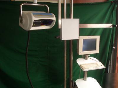
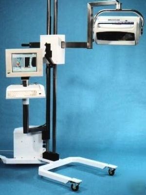
> Moor LD12 ir laser doppler imaging system MOORLD12IR
Moor LD12 ir laser doppler imaging system MOORLD12IR
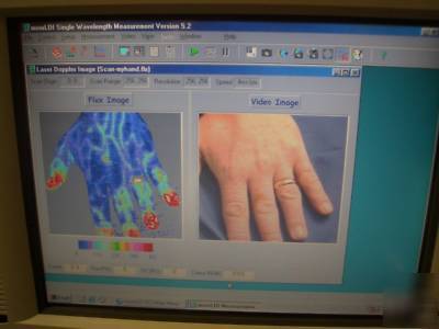
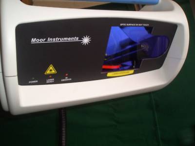
The Moor Laser Doppler Imager is a two dimensional laser scanning system that can image superfical blood perfusion on both large (full torso) and small tissue areas. The unit can be controlled directly or by a computer interface with results displayed using Windows-based software. A major application is burns assessment, but of course any area of skin blood flow can be scanned.
The moorLDI laser Doppler images the tissue surface and maps blood flow. A full resolution scan includes 65,536 individual laser Doppler measurements and these are displayed alongside a corresponding 'photo' image, as a colour coded image.
Image size up to 256x256 pixels (not interpolated) with 1 mm resolution
Image area - 5x5cm (at 20cm distance) to 50x50cm (at 100cm distance)
Scan time - 4,10 or 50ms per pixel (under 5 minutes for full scan)
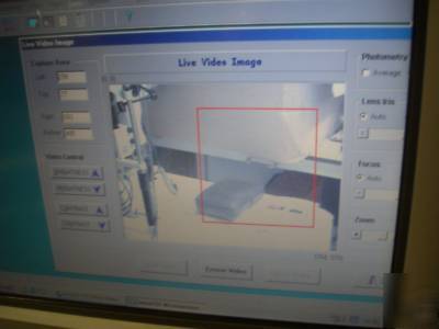
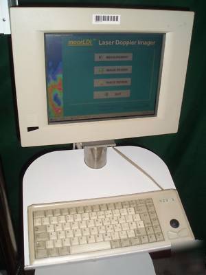
Windows-based software for control, processing & analysisis
Multiple patient & image file formats - moorLDI, ASCII, BMP,PCX, TIFF
The basic principle is that a low power laser light is used to illuminate the skin tissue, most of the light is reflected back by the static structures but some is reflected back by moving blood cells. The blood cells cause a Doppler shift in the wavelength of the scattered laser light which is proportional to the speed of the moving blood cells. The signal is processed by a microprocessor which can calculate the flux or blood flow. The tissue thickness measured is usually around 1mm, volume measure 1mm3, capillary diameter 10 m and velocities between 0.001 to 10mm/s. Laser Doppler Flowmeters use fibre optic cables placed on the surface of the skin to transmit and detect the laser light. The blood flow signal is displayed on the screen and shows changes in flow in time with the heart beat.
In Laser Doppler imaging, the laser light is directed via a moving mirror to scan a pattern across the tissue surface (see Fig 1). The reflected laser light is directed by the same moving mirror onto image detectors. The intensity fluctuations on the detector are processed to give an image of blood flow in the skin.